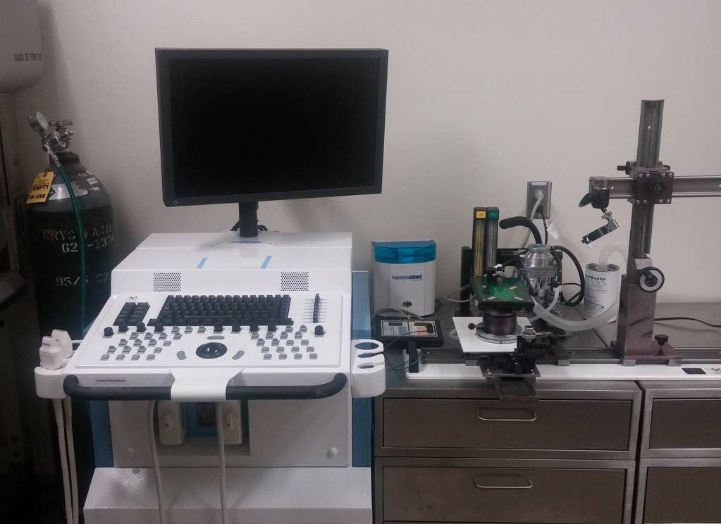
Echocardiography is a versatile and essential non-invasive tool for visualizing cardiovascular structures and evaluating cardiac function in mice. The UAPC maintains a state-of-the-art Visualsonics Vevo 2100, which offers near microscopic spatial resolution of ~30 microns and high temporal resolution up to 1000 frames per second. A comprehensive mouse echocardiography often includes:
-2D long- and short-axis cineloops of the heart
-M-mode images of the left ventricle and key structures
-Pulse-Wave Doppler recordings of aortic and trans-mitral valve velocities
-Tissue Doppler recordings of mitral annulus and left ventricle wall motion velocities
In the presence of wall motion abnormalities (i.e. after a myocardial infarction), more advanced analysis, including strain rate measurements, may be warranted.
Echocardiography is typically performed in anesthetized mice; however, echocardiography is possible in conscious mice. Training of mice in order to facilitate acclimation to the procedure may be required.
Image acquisition protocols and measurement can be customized based on investigator needs. For a complete list of echocardiography services and data output available see our Echo Request Form or Contact Us.


